Shoulder Muscles Diagram Labeled ~ The Shoulder Joint - Structure - Movement - TeachMeAnatomy. Learn the anatomy of the shoulder shoulder muscles: The shoulder muscles include skeletal muscles that are attached to the head of the humerus which performs various direct and indirect functions of the shoulder joints. 6 photos of the shoulder muscles labelled diagram. Muscles of the rotator cuff labeled on a sagittal mr slice. Around the shoulder, muscles in the back, neck, shoulder, chest and upper arm all work together to support.
Human anatomy diagrams show internal organs, cells, systems, conditions, symptoms and sickness information and/or tips for healthy living. Male anterior thoracic wall chest muscles labeled on white. Human muscle system functions diagram facts britannicacom. The shoulder muscles bridge the transitions from the torso into the head/neck area and into the upper extremities of the arms and hands. Chest muscles diagram anatomy shoulder anatomy shoulder muscle.
Top 10 Strongest Muscles in The Body | Pouted.com from www.pouted.com See below to view an image of the rotator cuff structure: The neck muscles and massive triangular muscles of the back stabilise the head and shoulders and permit a range of complex movements. Role of the shoulder muscles in controlling the glenohumeral joint and prevention of subacromial impingement in overhead. The shoulder is not a single joint, but a complex arrangement of bones, ligaments, muscles, and tendons that is bones of the shoulder girdle. Start studying shoulder muscles labeling. The shoulder muscles are associated with movements of the upper limb. Test your knowledge in our quiz about the shoulder muscles. Learn the anatomy of the shoulder shoulder muscles:
The shoulder muscle tissues can be readily injured and therefore being aware of the appropriate strategy is pretty significant when functioning out.
Supraspinatus, infraspinatus, ters minor,.et), using interactive animations and labeled diagrams. Mri of the shoulder : Posted on january 21, 2015 by admin. Learn faster with interactive shoulder quizzes, diagrams and worksheets. The muscular system is responsible for movement in collaboration with the nervous system to form impulses for motion. This diagram with labels depicts and explains the details of muscles on shoulder. Each anatomical structure was interactively labeled. Which are the shoulder muscles and where they are located? Although three ligaments protect and surround the shoulder joint, most of its stability comes from the powerful muscles and tendons of the rotator cuff. Human anatomy diagrams show internal organs, cells, systems, conditions, symptoms and sickness information and/or tips for healthy living. In this video we'll explore the muscles and functions of the shoulder girdle (pectoral girdle). The shoulder has about eight muscles that attach to the scapula, humerus, and clavicle. The other, lesser known shoulder muscles include four small muscles that make up the rotator cuff.
This diagram depicts shoulder muscle diagram. Labeled muscle diagram 10241878 diagram labeled muscle diagram 10241878 chart human anatomy diagrams and charts explained. Muscles also contribute to internal functions of the human body which include motion in the intestines and circulatory system. The clavicle scapula and humerus. When the nervous system signals the muscle to contract groups of muscles work together to move the skeleton.
Female Shoulder Muscles Diagram - Pin on Exercise : The human shoulder is made up of three bones ... from www.anatomynow.com Learn faster with interactive shoulder quizzes, diagrams and worksheets. Muscles of the rotator cuff labeled on a sagittal mr slice. They work closely with the shoulder girdle muscles to stabilize and move the shoulder. Which are the shoulder muscles and where they are located? Posted on january 21, 2015 by admin. Muscle charts of the human body for your. Learn vocabulary, terms and more with flashcards, games and other study tools. The anterior deltoid, the lateral deltoid, and the posterior deltoid.
Muscle charts of the human body for your.
The human shoulder is made up of three bones: The muscular system is responsible for movement in collaboration with the nervous system to form impulses for motion. Posted on january 21, 2015 by admin. Free access interactive and dynamic anatomical atlas. Labeled muscle diagram 10241878 diagram labeled muscle diagram 10241878 chart human anatomy diagrams and charts explained. Muscles also contribute to internal functions of the human body which include motion in the intestines and circulatory system. Muscle charts of the human body for your. I hope you find them as useful as i do. We have collected the best muscle diagram including arm, facial, neck, quadriceps, and shoulder muscle diagrams. This diagram depicts shoulder muscle diagram. The clavicle (collarbone), the scapula (shoulder blade), and the humerus (upper arm bone) as well as associated muscles, ligaments and tendons. The shoulder muscles include skeletal muscles that are attached to the head of the humerus which performs various direct and indirect functions of the shoulder joints. Muscle diagram of shoulder shoulder muscles diagram shoulder muscle diagrams diagram site.
These muscles form the outer shape of the shoulder and underarm. Help yourself in studying the anatomy of the body muscles with this set of free and printable human muscles diagrams labeled 2019. See below to view an image of the rotator cuff structure: This diagram depicts shoulder muscle diagram. Male anterior thoracic wall chest muscles labeled on white.
Why Am I Having Sharp Shoulder Pain? - Girls Gone Strong from www.girlsgonestrong.com Human muscles enable movement it is important to understand what they do in order to diagnose sports injuries and prescribe rehabilitation exercises. This diagram depicts shoulder muscle diagram. Test your knowledge in our quiz about the shoulder muscles. I hope you find them as useful as i do. The shoulder muscles are associated with movements of the upper limb. Exercise of this organ system is critical to prevent. Free access interactive and dynamic anatomical atlas. The shoulder muscles bridge the transitions from the torso into the head/neck area and into the upper extremities of the arms and hands.
The extrinsic muscles of the shoulder include trapezius, latissimus dorsi, levator scapulae, rhomboid major and rhomboid minor.
The rotator cuff is a complex and delicate structure of. Each anatomical structure was interactively labeled. Learn vocabulary, terms and more with flashcards, games and other study tools. 7 draw labelled diagram showing the relations of axillary nerve also supplies skin over lower half of deltoid (regimental badge area), therfore sensory. Muscle charts of the human body for your. Together these are known as the rotator cuff muscles. Which are the shoulder muscles and where they are located? Now label the diagram in your workbook! The shoulder is not a single joint, but a complex arrangement of bones, ligaments, muscles, and tendons that is bones of the shoulder girdle. The shoulder muscles include skeletal muscles that are attached to the head of the humerus which performs various direct and indirect functions of the shoulder joints. Ankle muscles diagram, back muscles diagram, chest muscles diagram, diagram of shoulder muscles and tendons, hip muscles diagram, knee muscles diagram, neck muscles diagram, rotator cuff muscles diagram, human muscles. The shoulder muscles are associated with movements of the upper limb. This diagram depicts shoulder muscle diagram.
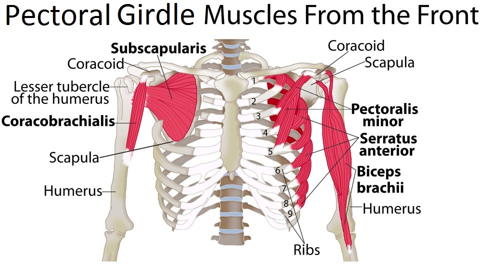
Male anterior thoracic wall chest muscles labeled on white shoulder muscles diagram. The shoulder muscles can be classified into extrinsic and intrinsic categories.
Share :
Post a Comment
for "Shoulder Muscles Diagram Labeled ~ The Shoulder Joint - Structure - Movement - TeachMeAnatomy"


Post a Comment for "Shoulder Muscles Diagram Labeled ~ The Shoulder Joint - Structure - Movement - TeachMeAnatomy"